GraphAbs
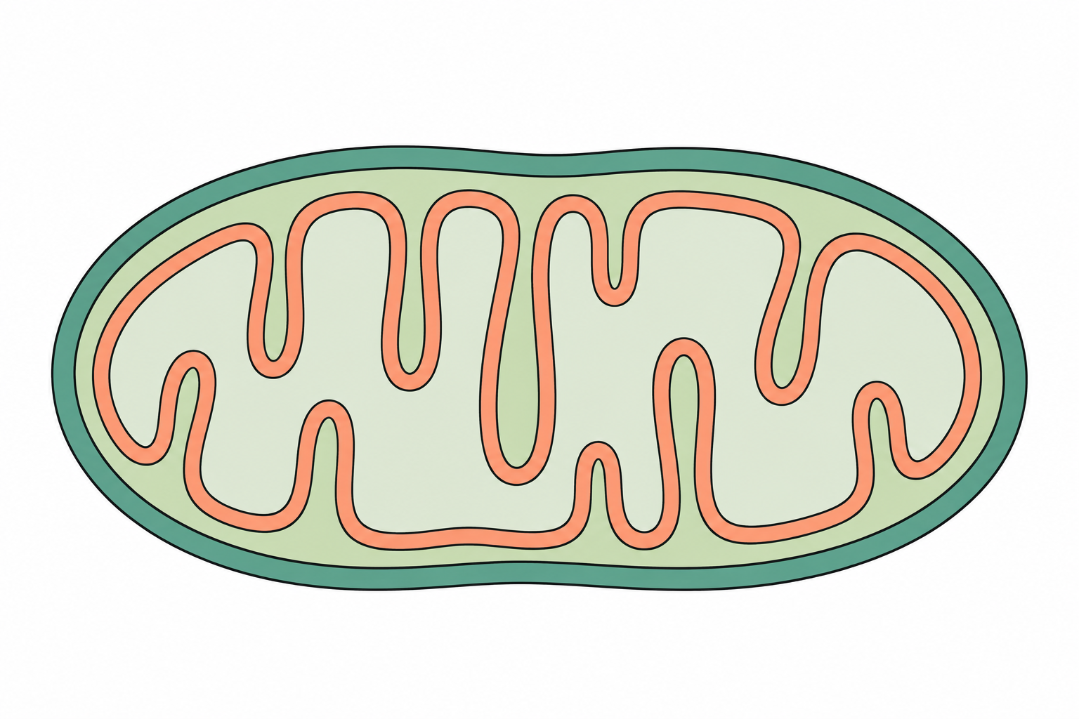
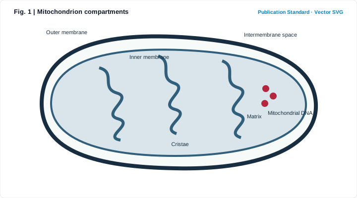
Create: Mitochondrion compartments
Edit the structures, labels and subject in the prompt, then generate an AI illustration. The example is an original vector schematic; AI output is a raster image. Review every structure and label before publication.
1. What is Mitochondrion compartments?
Scientific definition and academic background
A mitochondrion diagram highlights its double-membrane organization. The folds of the inner membrane create cristae, while the matrix contains enzymes and mitochondrial components.
The two membranes separate compartments with different roles in energy metabolism.
2. Key Visual Anatomy & Elements
Deconstruction of axes, visual geometries, and annotations
Clear Boundaries & Hierarchy
Distinguishes outer boundaries, internal compartments, and relative spatial layout.
Directional & Process Cues
Uses consistent arrow styles to indicate flow, sequence, or regulation without ambiguity.
Standardized Callout Labels
Aligned sans-serif labels sized to remain legible at journal single-column print widths.
Restrained Journal Palette
Clean white background with 3–5 muted hues that keep focus on scientific meaning.
3. Core Structure & Labeling Checklist
Key structures, reading order, and visual cues to specify before generating
- List the exact anatomical or conceptual structures required in Mitochondrion compartments so no critical label is omitted.
- Specify a clean white background and flat editorial vector style—avoid decorative 3D glare or clutter.
- Define the reading order (e.g., outer-to-inner, left-to-right, or numbered stages) for clear comprehension.
- Verify every label spelling, arrow direction, and scientific relationship before manuscript submission.
4. Reusable Scientific Illustration Prompt
Copy or load this structured prompt into the studio and customize the labels
Mitochondrion compartments. A mitochondrion diagram highlights its double-membrane organization. The folds of the inner membrane create cristae, while the matrix contains enzymes and mitochondrial components. Include these labels: Outer membrane, Inner membrane, Cristae, Intermembrane space, Matrix, Mitochondrial DNA. White background; restrained navy, crimson and sage scientific palette. Simplified schematic, not to scale.
5. Scientific Insights & Publication Legend
Hypothesis addressed and a ready-to-use Nature/Cell figure legend template
Core Scientific Question:
How can a clear, unambiguous Mitochondrion compartments schematic communicate core structures and processes at a glance?
Interpretation Guide:
Condensing dense background text into a structured visual anchor helps reviewers grasp your model immediately and pairs cleanly with data panels in multi-panel figures.
Standard Journal Figure Legend Template:
6. Peer Review Solutions & Pitfall Prevention
Overcoming reviewer critiques and eliminating common desk-rejection traps
Reviewer notes: "The text description is dense; a clear overview schematic is needed."
Generates a structured publication draft from this template with in-place regional refinement.
Reviewer notes: "A specific label or directional arrow in the schematic needs correction."
Circle the region in the studio to revise only that area while preserving the rest of the layout.
7. Reviewer Checklist & Journal Formatting Specs
Four indispensable pre-submission quality checks to ensure seamless editorial pass
Column Width
Nature single column (89 mm) or full width (183 mm), max height 220 mm
Typography
Arial / Helvetica, 8–9 pt for headings, 7–8 pt for labels
Palettes
Restrained academic palette (Navy, Ochre, Sage), avoiding neon saturations
Resolution
300–600 DPI print resolution, or assemble with target journal widths in TIFF / SVG
Cell biology note
Mitochondrial size, number, and cristae shape vary with cell type and metabolic state. This generic drawing should not be read as a micrograph.